
We provide end-to-end histopathology and precision medicine services from sample to score for clinical trials and clinical research.
AcelaBio’s pathology workflow is fully digital and includes automated sample tracking with a documented audit trail from sample receipt to sign-out. Our team brings expertise in providing clinical research services to pharmaceutical, biotech, and academic institutions.
Take a tour of our state-of-the-art commercial lab.
AcelaBio offers:
Automated sample tracking with documented audit trail from sample receipt to sign-out.
Services for clinical trials, pharmaceutical research, biotechnology, government agencies, and the investigator community.
Proven technology, quality management systems, and access to leading doctors and scientists

Accessioning
Accessioning
Cross-referencing of sample identifiers with shipping manifest or requisition form
Confirmation of completeness of sample-associated meta-data
Registration of sample in laboratory information system

Processing
Processing
Fully automated process to dehydrate, clear, and infiltrate the tissue
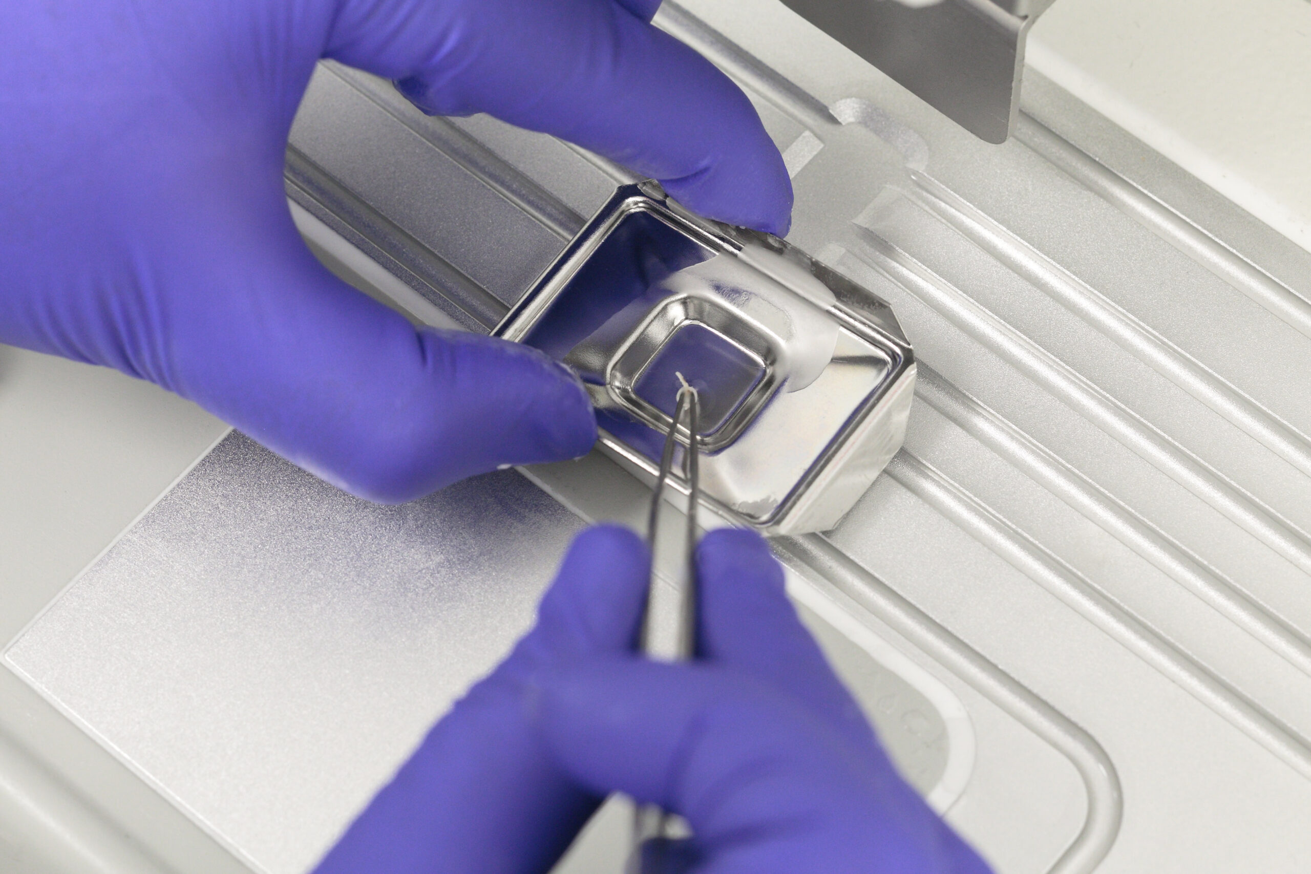
Embedding
Embedding
Embedding of processed tissue into a wax block
Ensuring that tissue is oriented in a correct way for downstream microtomy

Sectioning
Sectioning
Microtomy with sectioning of the wax block to cut sections of typically 4 µm thickness
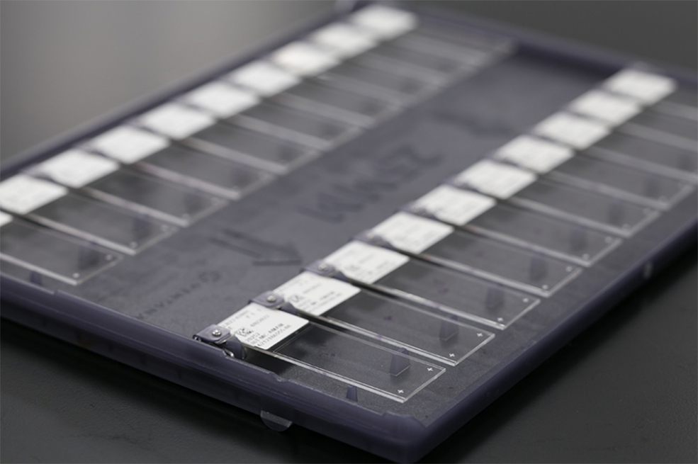
Tissue sections are subsequently placed on positively charged glass slides

Hematoxylin & Eosin
Hematoxylin & Eosin
Automated staining of tissue on a glass slide with hematoxylin and eosin
Hematoxylin stains cell nuclei a purplish blue
Eosin stains the extracellular matrix and cytoplasm pink

Special Stains
Special Stains
Automated special staining of tissue on a glass slide, such as with Masson Trichrome
Immunostaining
Immunostaining
Automated staining with antibodies linked to chromogens to identify a certain target
Single or multiplex chromogenic immunohistochemistry (IHC)

Assay Development
Assay Development
Development of a new staining protocol, typically using immunohistochemistry
The starting point can be a known target, an antibody, or an in vitro diagnostic (IVD)

Antibody Optimization and Validation
Antibody Optimization and Validation
Process of characterizing and validating antibodies for the identification of biomarker targets
Testing out different dilutions, buffers, and reaction kits

Whole Slide Image Scanning
Whole Slide Image Scanning
Digital pathology using 510(k) approved scanner to scan whole glass slides up to 40X
Digitized slides can then be transferred to an image management system for pathologist review
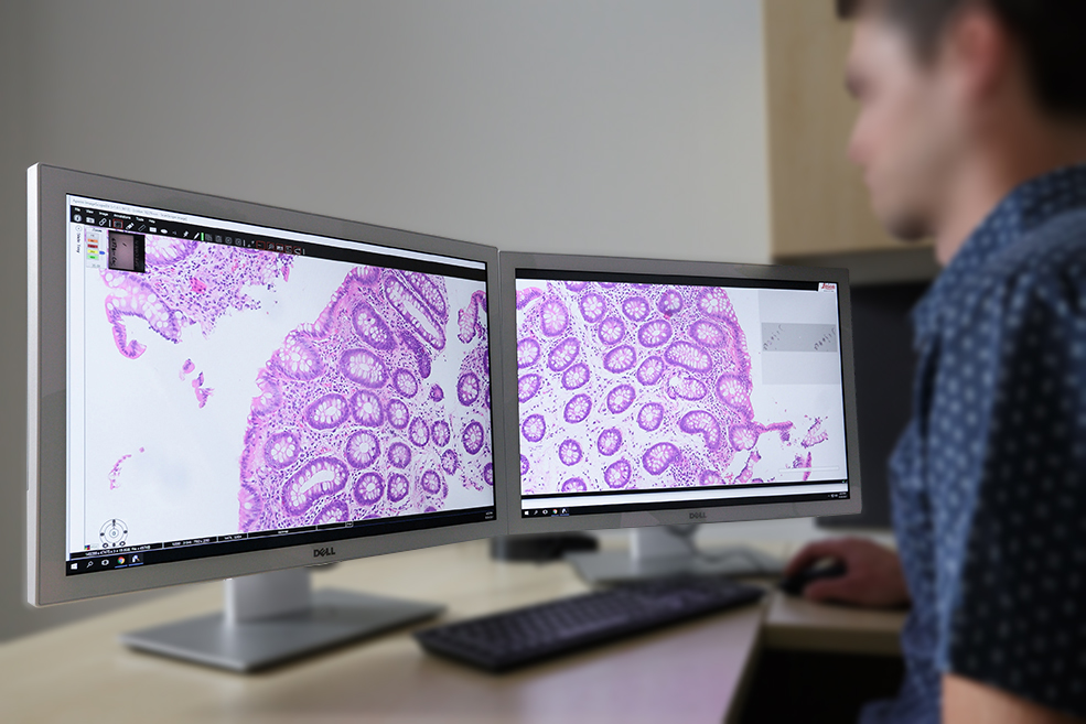
Pathology Review
Pathology Review
Support assay development as it relates to ensuring staining pattern is correct
Identification and annotation of tissue areas, structures, and cells to support the development of algorithms for quantitative digital image analysis
Review of clinical cases, which may include disease activity scoring and diagnosis

Data Management
Data Management
Delivery of consistent data & imagery through automated laboratory information systems

Sample Logistics
Sample Logistics
Preparation and shipment of blocks and slides to national and international locations
Storage & Archiving
Storage & Archiving
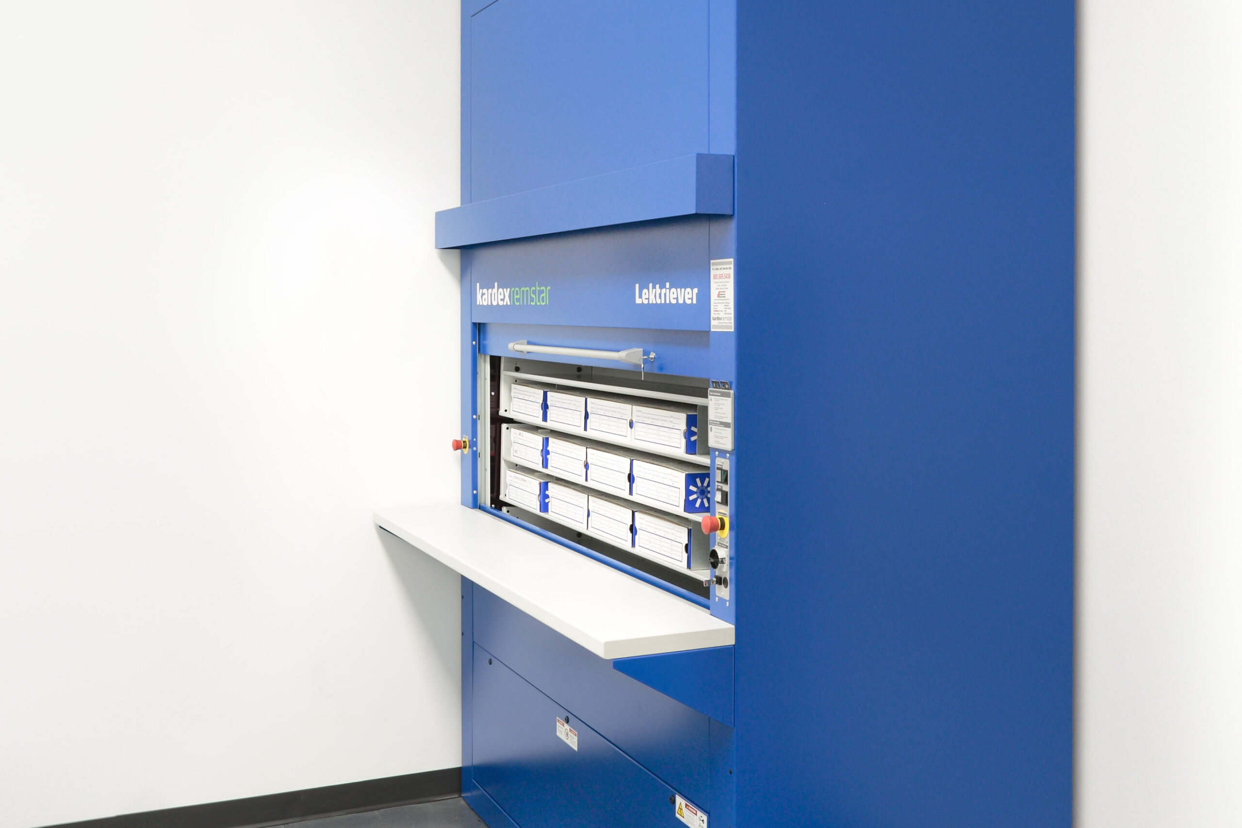
Semi-automated archival system of blocks and slides in temperature & humidity-controlled environment

Consulting
Consulting
Implementation of histopathology services in multicenter, global clinical trials
Selection of biomarker targets based on mechanism of action and technical capabilities
Project Management
Project Management
Operational experts ensure projects are delivered on time within budget

In Situ Hybridization (ISH)
In Situ Hybridization (ISH)
Use of specific probes to identify target gene transcripts in tissue using RNAscope

Clinical Trial Support
Clinical Trial Support
AcelaBio works in partnership with Alimentiv, a leading full-service niche CRO specialized in gastroenterology, to provide an end-to-end clinical study management solution that ranges from early trial design to complete execution of a clinical trial.
**For GI Services Only

Digital Image Analysis*
Digital Image Analysis*
Quantitative digital image analysis using machine learning and AI techniques
Custom algorithm development with oversight of expert pathologists
Generate quantitative, reproducible data on whole slide images or specific regions of interest
*For Research Use Only (not for use in diagnostic procedures)


